
“Congrats! You’re gonna be Mom and Dad!” OK, give you a second to burst into tears, then hug, kiss, and touch the belly. Then, confusion and worry kick in. What to prepare next? Well, clothes or gender reveal party later, move the medical checking ahead. After all, security is the most important.
Among all the tests during the pregnancy, the color Doppler test is the most fundamental. This blog will explore the details of the color Doppler test during the prenatal period.
Table of Contents
What is Color Doppler Ultrasound in Prenatal Test
Color Doppler ultrasound, abbreviated as CDUS, is a medical imaging examination method that uses ultrasound technology.
By transmitting high-frequency sound waves into the body and recording the signals reflected between the sound waves and tissues, it obtains imaging information about the fetus.
It can provide accurate information on fetal development, placental function, amniotic fluid volume, and other important indicators, helping doctors assess the health status of the fetus and identify any potential abnormalities. Therefore, it is of great significance for screening and diagnosing complications during pregnancy and fetal development issues.
The Differences Among Prenatal Ultrasound
Prenatal ultrasound concludes with 2D, 3D, 4D, and color ultrasound.
2D Ultrasound & Color Doppler Ultrasound
Color Doppler ultrasound, also known as an upgraded type of 2D color Doppler ultrasound, is an advanced imaging technique that combines the principles of Doppler ultrasound with color imaging technology. It enhances the traditional grayscale ultrasound images by overlaying color signals onto them, typically represented in shades of red and blue.
This color enhancement allows for better visualization of blood flow direction and velocity, providing valuable insights into vascular dynamics. Compared to conventional B-mode ultrasound, color Doppler ultrasound offers superior clarity and detail.
In addition to its enhanced imaging capabilities, color Doppler ultrasound shares similarities with conventional B-mode ultrasound in that both modalities display cross-sectional images. However, the incorporation of color Doppler technology adds an extra dimension to the imaging process, enabling clinicians to assess blood flow patterns and vascular abnormalities with greater precision.
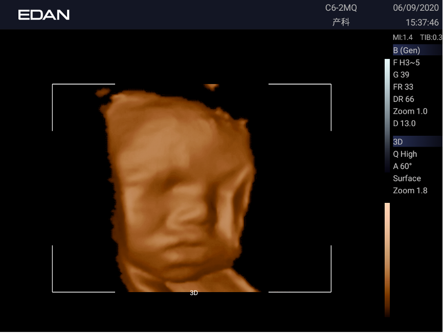
3D & 4D Ultrasound
Building upon the foundation of color Doppler ultrasound, three-dimensional (3D) and four-dimensional (4D) ultrasound techniques further expand the diagnostic capabilities in prenatal imaging.
3D ultrasound provides volumetric imaging capabilities, allowing for multi-angle observation of lesions and improved spatial visualization. On the other hand, 4D ultrasound introduces the element of “Time” to the imaging process, enabling dynamic visualization of fetal movements and anatomical structures over a period of time.
Is 3D or 4D Ultrasound More Superior?
While 3D and 4D ultrasound offer valuable insights into fetal development and maternal health, it’s essential to note that conventional 2D ultrasound remains the cornerstone of prenatal imaging.
During routine fetal ultrasound examinations, clinicians primarily utilize 2D ultrasound to comprehensively evaluate fetal anatomy and monitor fetal well-being.
The integration of 3D and 4D ultrasound techniques is typically reserved for specific clinical scenarios or diagnostic needs, serving as complementary imaging modalities to enhance diagnostic accuracy and provide additional clinical information.
When to be Prescribed for Prenatal Ultrasound
First Trimester Scan (6-8 weeks): This initial scan aims to confirm intrauterine pregnancy, rule out ectopic pregnancy, determine the number of fetuses (singleton, twins, or multiples), assess embryo implantation, detect fetal cardiac activity, and verify the consistency between gestational age and the last menstrual period.
Nuchal Translucency (NT) Scan (11-13 weeks + 6 days): The NT scan primarily measures the thickness of the fetal nuchal translucency, aiming to identify potential neural tube defects, chromosomal abnormalities, or cardiovascular anomalies.
Second Trimester Anomaly Scan (22-24 weeks) and Third Trimester Scan (29-31 weeks): These scans are systematic screenings aimed at detecting severe fetal abnormalities, evaluating fetal growth, assessing amniotic fluid volume, and examining fetal anatomy comprehensively.
Late Pregnancy Scan (37-41 weeks): Conducted in the late stages of pregnancy, this scan focuses on assessing fetal development, amniotic fluid levels, fetal position, and placental position. It evaluates fetal size, amniotic fluid volume, placental maturity, and umbilical artery blood flow.
Pregnancy Ultrasound Test to the High-Risk Group
For the group who are considered high-risk, it means that the mother or the fetus might be more likely than usual to develop health problems before or during pregnancy.
The Reasons for High-Risk Pregnancy
High-risk pregnancy results from various factors:
- Age: Certain maternal ages (being over 35 or under 17 when pregnant) pose greater risks, including miscarriage, preterm birth, and chromosomal abnormalities. Ultrasound monitoring can help doctors identify potential issues early and take appropriate measures.
- Chronic Diseases: Pregnant women with chronic conditions such as diabetes and hypertension are considered high-risk due to the likelihood of complications. Color Doppler ultrasound can detect fetal growth issues early, providing a basis for effective management.
- Multiple Pregnancies: Women carrying twins, triplets, or more face higher risks, including preterm birth and low birth weight. Ultrasound monitoring can assess the health status of each fetus in detail, ensuring they receive adequate nutrition and care.
- Pregnancy Complications: Conditions like preeclampsia and placental abruption can threaten the lives of both mother and baby. Ultrasound monitoring is crucial for timely understanding of fetal development and placenta position, aiding in prompt and effective intervention.
Importance of Ultrasound Test for the High-Risk Pregnancy Group
- Early Screening: Ultrasound monitoring in early pregnancy allows for the screening of potential issues such as fetal anomalies, congenital heart defects, and other abnormalities. This information forms the basis for physicians to develop personalized treatment plans.
- Growth and Development Monitoring: Regular ultrasound examinations provide comprehensive insights into fetal growth and development, ensuring that the fetus receives appropriate nutrition and care at different stages of pregnancy.
- Complication Monitoring: For pregnant individuals with pre-existing conditions or complications, ultrasound monitoring aids in the timely detection and monitoring of changes in their condition. This enables healthcare providers to take appropriate treatment measures to mitigate risks.
- Management of Multiple Pregnancies: Ultrasound monitoring plays a crucial role in the management of multiple pregnancies. Physicians can monitor the development of each fetus, detect potential complications early, and intervene as necessary to ensure the best outcomes for both the mother and the babies.
Diagnostic Ultrasound System from EDAN
EDA, as the experienced provider of phenomenal medical devices and health solutions, rolled out a series of innovative ultrasound devices to safeguard the health of mothers and babies.
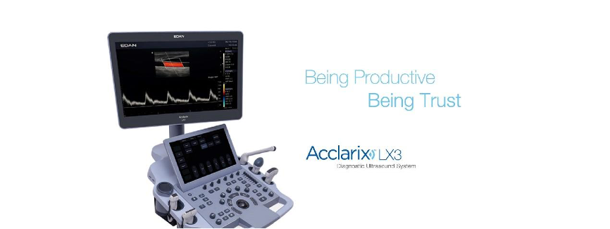
Acclarix LX3 diagnostic ultrasound system, for example, enjoys great applause.
As a ground-breaking ultrasound system with sovereign IPRs, it excels in design and performance. The compact body, innovative technology, and intelligent workflow make it particularly effective in handling complex ultrasound environments.
Acclarix LX3 features
- Fully activated five-probe slots
- A wide array of probes
- Self-guided teaching software
- Various imaging technologies
They significantly enhance physicians’ work efficiency. Additionally, its superior image quality is invaluable for early screening and diagnosis, earning it high recognition.
Conclusion
Regular screening and checking are by no means one of the basic safeguards for mothers and babies. And the higher accuracy and efficiency hinge on the daily advancing diagnostic devices.
So, to see further healthcare solutions, you can visit EDAN.